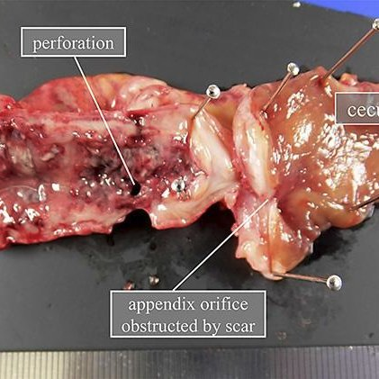
Gross Examination
Gross examination in pathology refers to the initial visual inspection and examination of tissue specimens or surgical specimens without the aid of a microscope. This examination is typically conducted by pathologists or pathology assistants. The purpose of gross examination is to identify any gross abnormalities, lesions, or other features that may provide insight into the underlying disease process.
Specimen Reception: The specimen is received from the surgical or clinical team and is labeled with relevant patient information, such as name, medical record number, date, and site of the specimen.
 Stock Photo ID: 2217987715 Biopsy samples being processed at the pathology laboratory to be embedded in paraffin for analysis.
Description: The pathologist or pathology assistant begins by describing the external features of the specimen, including its size, shape, color, and any abnormalities that are visible to the naked eye. This description is recorded in the pathology report.
 A Pathologist Assistant in Histology performs a gross examination of a tissue specimen that has come from the operating room.
Measurement: The dimensions of the specimen are measured using rulers or calipers. This is important for documenting the size of lesions or abnormalities, as well as determining the adequacy of the specimen for diagnostic purposes.
Sectioning: The specimen may be sectioned using a scalpel or other cutting instrument to expose internal structures or lesions. The pathologist may choose specific areas for further examination based on clinical history, imaging studies, or gross findings.
Sampling: Tissue samples are taken from various regions of the specimen for further processing and microscopic examination. The selection of sampling sites is guided by the gross examination findings and the diagnostic question at hand.
 Orientation during Sampling in Surgical Pathology, Gross pathology, IZAK B. DIMENSTEIN, MD, PHD, HT (ASCP)FacebookGooglePlus
Photography: Photographs may be taken to document the gross appearance of the specimen before it is processed further. These photographs can be included in the patient's medical record or used for teaching purposes.
Dissection: In some cases, particularly with complex specimens, further dissection may be necessary to fully examine internal structures or to isolate specific areas of interest for sampling.
Fixation: After gross examination and sampling are complete, the remaining specimen is typically fixed in formalin to preserve tissue structure for subsequent processing and histological examination.
Documentation: All findings from the gross examination, including descriptions, measurements, sampling sites, and any relevant clinical information, are documented in the pathology report.
Overall, gross examination plays a crucial role in the accurate diagnosis and management of diseases by providing valuable information about the macroscopic features of tissue specimens.
References:
- Mohan, H. (2013) Pathology practical book. 3rd edn. New Delhi: Jaypee Brothers Medical Publishers.
- Histopathology sampling guide for Livestock (n.d.) Available at: https://www.agric.wa.gov.au/livestock-biosecurity/histopathology-sampling-guide-livestock (Accessed: 28 March 2024).
- Prematilleke, I., Ratnayake, P. and Sosai,C. (2021) 'General guidelines for handling of tissue specimens for histopathological examination' in Amarathunga,P., Lokuhetty, D. and Prematilleke, I. (ed.) National Guidelines in Histopathology Collection, Handling and Transport of Surgical Specimens. , Sri Lanka: Ministry of Health, Sri Lanka, pp. 2-18.


Comments
Post a Comment