An Analysis of Virtual Microscopy: At University of Kelaniya

A novel approach has been implemented by the University of Kelaniya to engage students in the intriguing field of microscopic investigation. This blog post delves into the immersive experience of a University of Kelaniya Virtual Microscope Session.
The following specimens were examined.
01.Hair Follicle

Under a microscope, a hair follicle is a small, tube-like structure embedded in the skin. Among its multiple layers is the connective tissue that surrounds the hair bulb and the inner and outer root sheaths.
The first two layers of skin, the dermis and epidermis, are home to hair follicles.
By observation, one can learn about the follicle's thickness, pigment distribution, developmental stage, and any anomalies such as infection or inflammation.
02.Skin Under the Microscope

The stratified squamous epithelium that makes up the epidermis. The uppermost layer, called keratin, is formed by the stratified epithelium. Keratinocytes, which produce keratin, make up the majority of the outermost layer.
Beneath the epidermis is a layer of tissue called the dermis, which is composed of glands, hair follicles, blood vessels, and sweat and sebaceous glands.
03.Apocrine gland

Apocrine glands resemble larger, more deeply stained structures than eccrine glands when examined under a microscope. They are surrounded by myoepithelial cells and typically have a larger lumen. Apocrine glands also exhibit decapitation secretion, which is the loss of the apical portion of the cell during secretion.
04.Pyogenic Granuloma - benign vascular lesions

A common benign vascular disease known as pyrogenic granuloma or lobular capillary hemangioma is characterized by a rapid capillary expansion. Under a microscope, it often looks like a mass of densely vascularized tissue with numerous tiny endothelial-lined blood vessels. These veins often have a lobular shape and may be surrounded by a fibrous stroma.
Pyogenic granuloma is a tumor that does not contain pus and is not contagious. Instead, it is believed to be a reactive lesion caused by stress or changes in hormones. Despite their tendency to bleed readily, pyogenic granulomas are benign; however, they may require surgery or other procedures for treatment.
Appears under a microscope as enlarged nuclei, overlapped nuclei, irregularly shaped membranes, and somewhat transparent membranes. Round structures found in the nucleus are known as nuclear inclusions.
05. Fat vacuoles

Under a microscope, an oval or circular structure filled with lipid droplets is called a fat vacuole. It would typically have a distinct boundary and seem translucent. Depending on the techniques used, the staining's extent may change. Cells that store fat and often contain fat vacuoles include adipose tissue cells and hepatocytes in the liver.
06. Parasite - Appendicitis

Enterobius vermicularis, better known as the pinworm, is one parasite that can result in appendicitis. Pinworms (Enterobius vermicularis) are small, thread-like worms with a tapered end that look like tiny
worms under a microscope. frequently in pediatric patients with appendicitis.
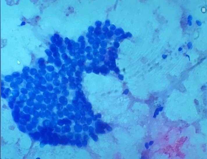
07. Cytology Sample- Peritoneum Fluid

Gramme stain is used in peritoneal fluid cytology to highlight the bacterial cell walls based on their stain-losing or stain-retaining capabilities. The purpose of this procedure is to search the sample for Gram-positive bacteria. Gram-positive purple bacteria keep the stain under a microscope.
08. Lymph Node and Capsule of the Lymph node

Under a microscope, the lymph node shows multiple distinct regions, such as the medulla, cortex, and paracortex.
A lymph node's capsule is visible as a thin layer of connective tissue encircling the node's outer edge.
In addition to carrying blood vessels and nerves that supply the node, it offers structural support. The capsule is essential to the preservation of the lymph node's structure and functionality.
Sub Capsular Sinus of the Lymph Nodes (empty space)

the separation between the capsule and the lymph node parenchyma. This sinus, which is lined with specialized cells, is the first location where lymph enters the lymph node. It acts as a filter, collecting foreign particles and antigens for immune cells to analyze and respond to appropriately.
Granuloma of Lymph Nodes

often notice a ring of lymphocytes surrounding a collection of immune cells, primarily macrophages. Compact clusters or nodules are common forms for these structures, though they can vary in size and shape.
Granulomas are a symptom of chronic inflammation and can be caused by a range of infectious and non-infectious agents, including bacteria, fungi, and foreign substances.
(All Images in this blog post were obtained from virtual microscope session during the site visit to the Department of pathology, Faculty of Medicine, University of Kelaniya.)
Related: "Pathology Outlines"
Click Here to view many related microscope slides virtually online.
References:
- Biology of the Hair Follicle: The Basics, Karoline Krause, MD, and Kerstin Foitzik, MD, Available at: https://scmsjournal.com/wp-content/uploads/2016/08/v25i1-Kraus.pdf
- Andrzej Slominski, Jacobo Wortsman, Neuroendocrinology of the Skin, Endocrine Reviews, Volume 21, Issue 5, 1 October 2000, Pages 457–487, https://doi.org/10.1210/edrv.21.5.0410
- Willard-Mack CL. Normal Structure, Function, and Histology of Lymph Nodes. Toxicologic Pathology. 2006;34(5):409-424. doi:10.1080/01926230600867727
- Postoperative intraperitoneal adhesion pathophysiology, J‐J. Duron, Journal: Colorectal Disease, 2007, Volume 9, Number s2, Page 14, DOI: 10.1111/j.1463-1318.2007.01343.x
Comments
Post a Comment